Draw A Labelled Diagram Showing The Structure Of A Neuron Biology

FileNeuron1.jpg Simple English Wikipedia, the free encyclopedia
The central nervous system ( CNS) consists of the brain and the spinal cord. It is in the CNS that all of the analysis of information takes place. The peripheral nervous system ( PNS ), which consists of the neurons and parts of neurons found outside of the CNS, includes sensory neurons and motor neurons.
Nerve Tissue SEER Training
The neuron (or nerve cell) is the functional unit of both the central nervous system (CNS) and the peripheral nervous system (PNS). The basic functions of neurons can be summarized into three main tasks: receiving signals, integrating these signals and transmitting the signals to target cells and organs.
Neurons The crazy wires in our body. Doc Jana
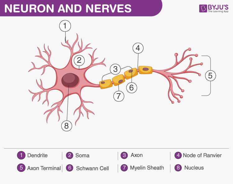
Choose the correct names for the parts of the neuron. (6) This neuron part receives messages from other neurons. (7) This neuron part sends on messages to other neurons. (8) This neuron part gives messages to muscle tissue. (9) This neuron part processes incoming messages. (10) This neuron part contains instructions for making proteins that the.
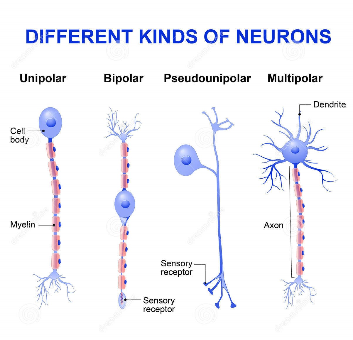
The Neuron External Structure and Classification Interactive
An Easy Guide to Neuron Anatomy with Diagrams By Olivia Guy-Evans, MSc Updated on November 9, 2023 Reviewed by Saul Mcleod, PhD Neurons are the information processing units of the brain responsible for sending, receiving, and transmitting electrochemical signals throughout the body.
What does a neuron look like? Biology Questions
3. How to Draw a Neuron Diagram To learn about the structure of the neurons, the students can use a neuron labeled diagram. The students may follow these steps to make their neuron diagram, but the process is complex: 3.1 How to Draw a Neuron Diagram from Sketch Step 1: First, the students need to draw a circle. Based on it, they need to draw a.
neural circuit diagram
Neuron Anatomy Activity The parts of the neuron have been labeled. Your challenge is to write the correct name for each part and explain what it does. If you need some help, visit the web article listed below. Neuron Anatomy Activity Synapses: Send electrical impulses to neighboring neurons.

What Is a Neuron? Diagrams, Types, Function, and More
An Easy Guide to Neuron Anatomy with Diagrams Anatomy Types Function Research Takeaway Neurons, also known as nerve cells, send and receive signals from your brain. While neurons have a lot.

Draw A Labelled Diagram Showing The Structure Of A Neuron Biology
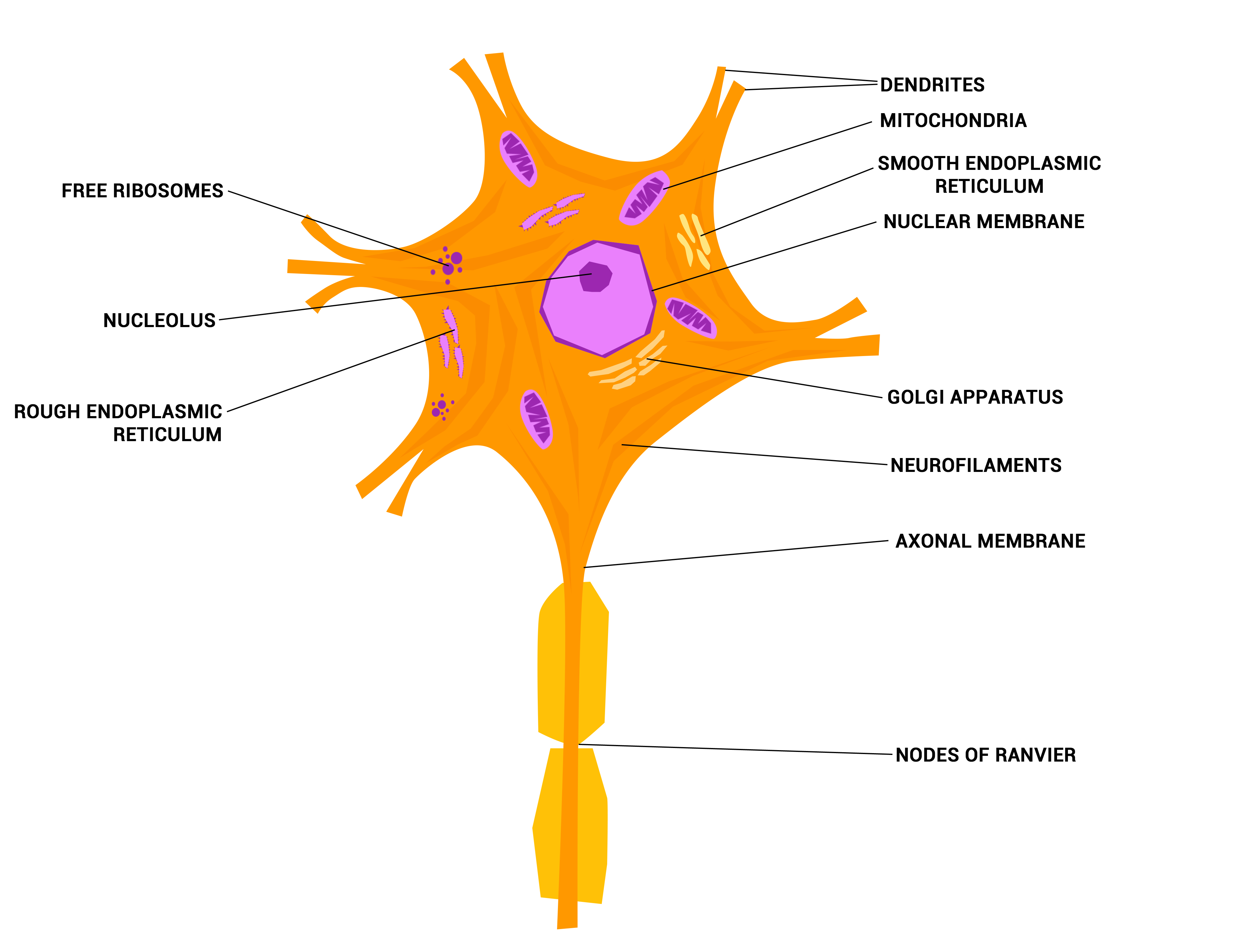
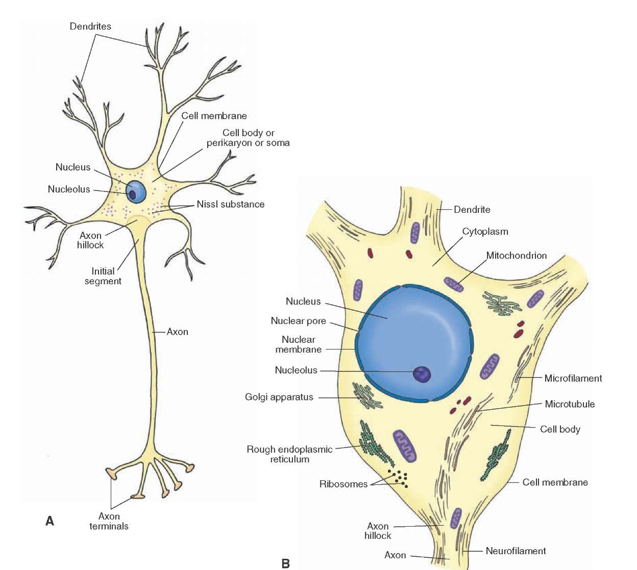
Parts of a Neuron and Their Functions with Labelled Diagram Parts of a Neuron and Their Function Neurons or nerve cells are the basic building blocks or units of the nervous system. Nearly 86 billion neurons work co-ordinately within the nervous system to keep the body organized.
Nerve Cell Diagram Labeled ClipArt Best
Diagram Of Neuron with Labels Here is the description of human neuron along with the diagram of the neuron and their parts. The neuron is a specialized and individual cell, which is also known as the nerve cell. A group of neurons forms a nerve.
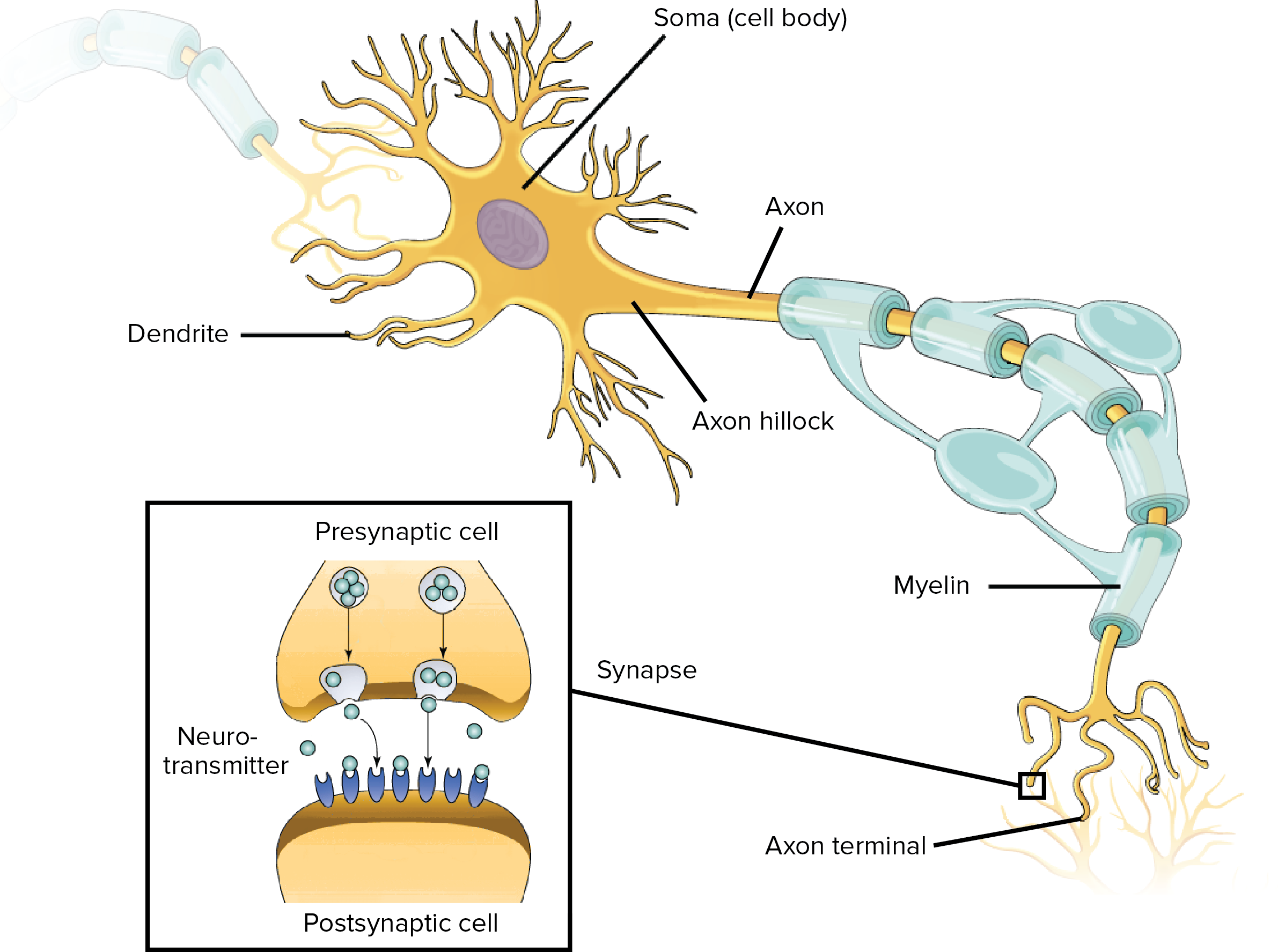
A Junction at Which a Neuron Meets Another Cell
A diagram of a neuron also known as the nerve cell is useful as a visual tool to illustrate the various components of the neuron. It also helps us to understand the functions of the neuron. This article incorporates a well-labeled diagram and a brief description of the components of a neuron highlighting their functions. Table of Content
Histology of the Nervous System (The Neuron) Part 1
Neuron Labeling Neuroglia Labeling with Google Slides Save paper by assigning labeling worksheets on Google Classroom. Instead of writing in labels, students drag and drop the labels to the appropriate area on the image. The labeling focuses on the neuron and supporting neuroglia (or glial) cells.
Structure and types of neuron (The nervous tissue) Online Science Notes
Cerebellum - molecular, Purkinje, granular layers. Peripheral nerves - epineurium, perineurium, endoneurium. This article will explain the histology of neurons, providing you with information about their structure, types, and clinical relevance. It will also cover briefly the histological layers of the central and peripheral nervous systems.
A diagram of a neuron and its functions. a study in chartreuse
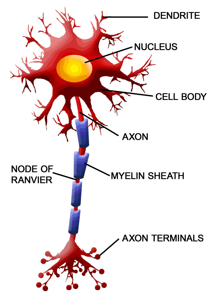
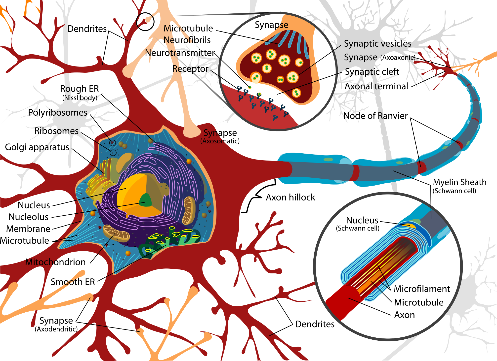
In the neuron drawing shown below, the neuron is labeled to show structures such as the cell body, axons, dendrites, and synapses. A detailed diagram of a neuron, showing the synapse, cell body.
neuron diagram labeled
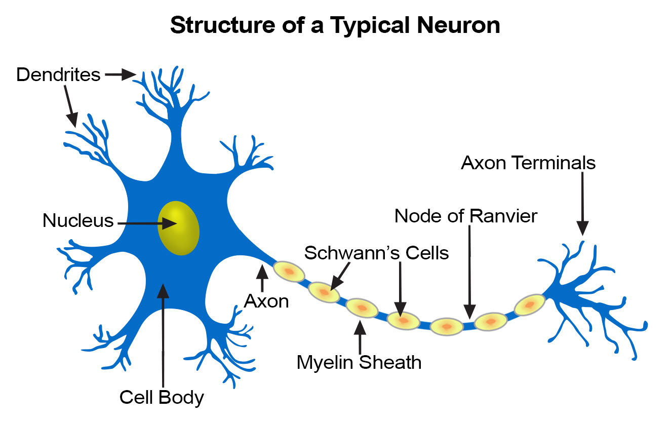
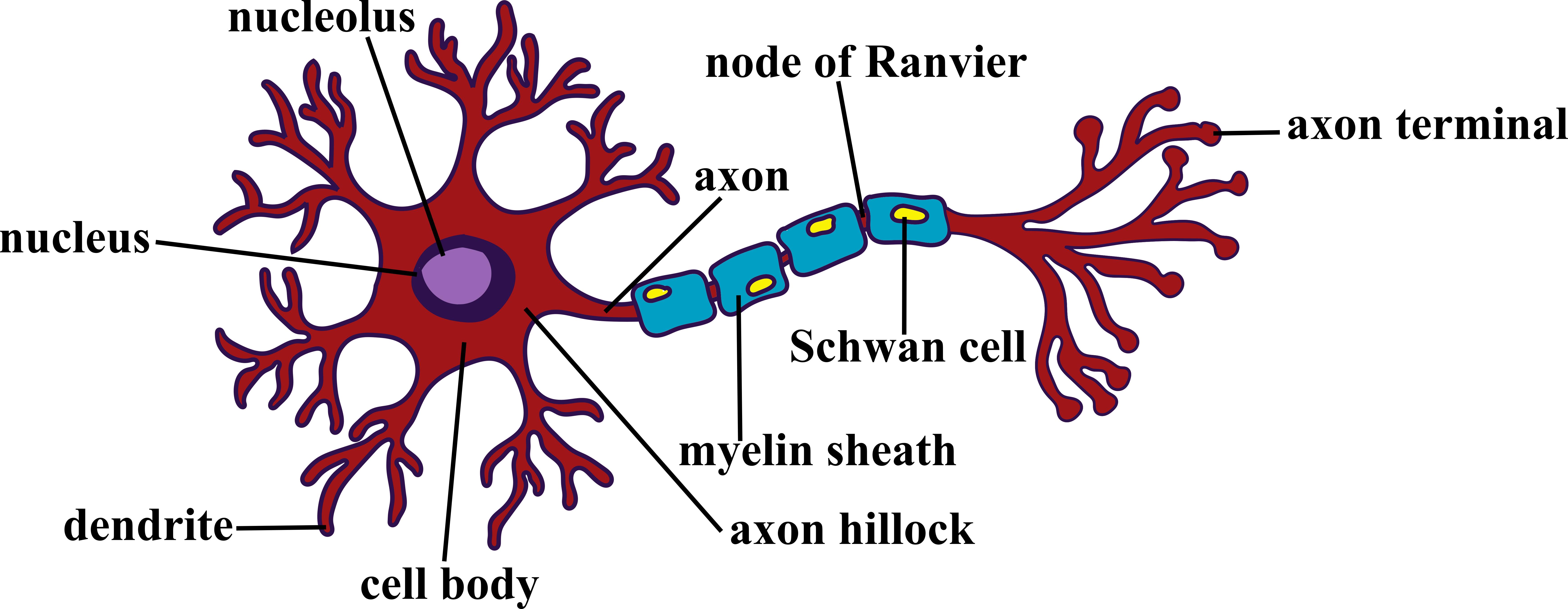
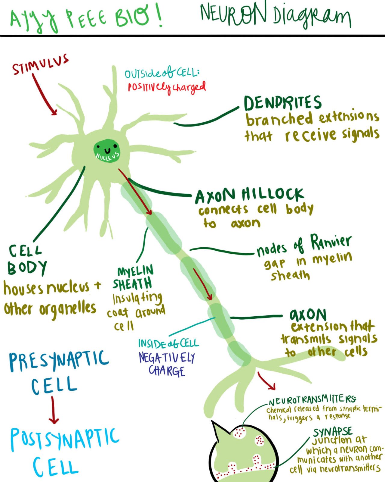
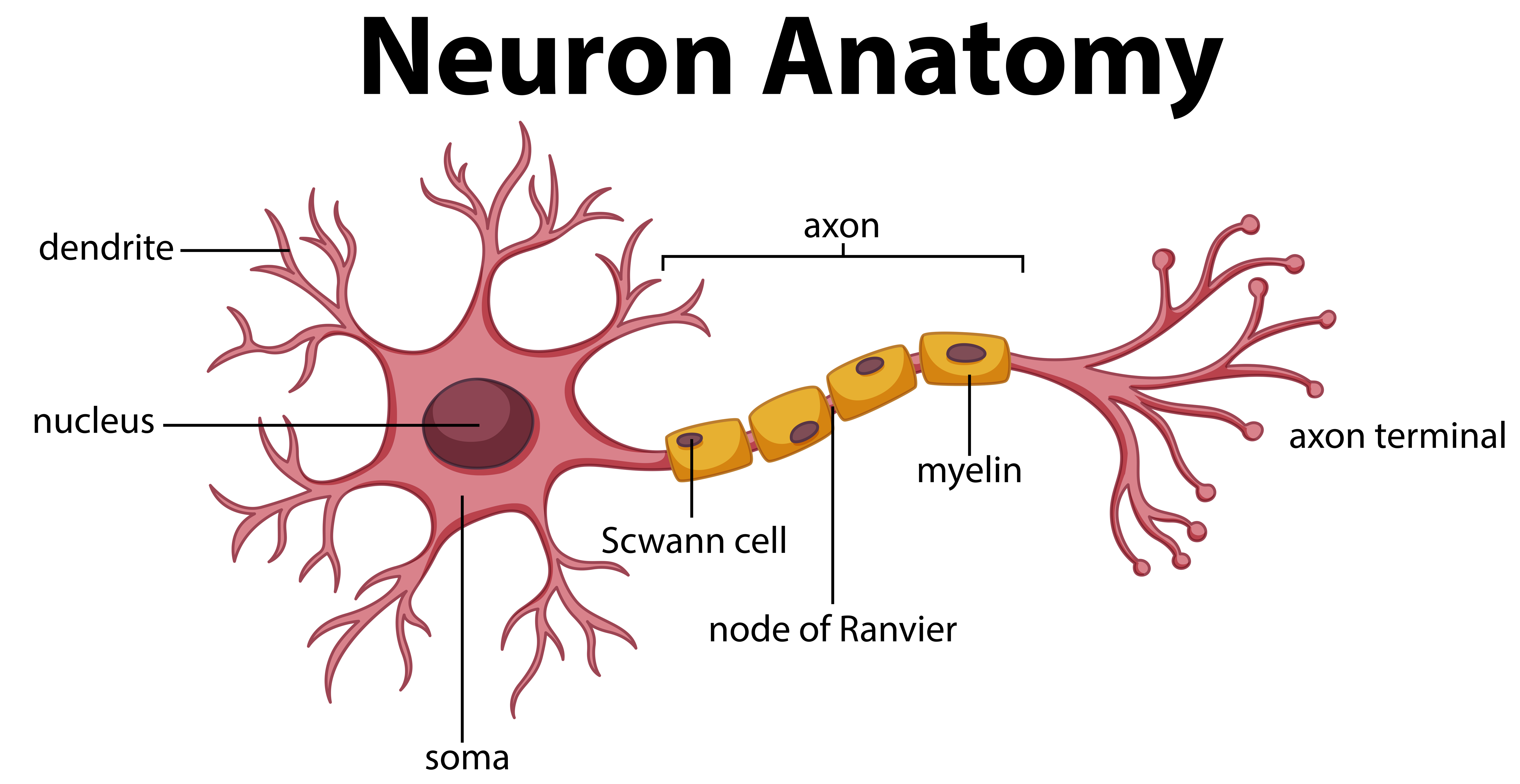
Dendrites The branched extensions of a neuron that receive input from other neurons. Myelin Fatty substance that forms a sheathlike covering around axons. Nodes of Ranvier Gaps between myelin sheaths on the axon of a neuron. Nucleus The cell organelle that contains nearly all the cell's DNA. Schwann cells

Anatomy Of A Neuron ANATOMY
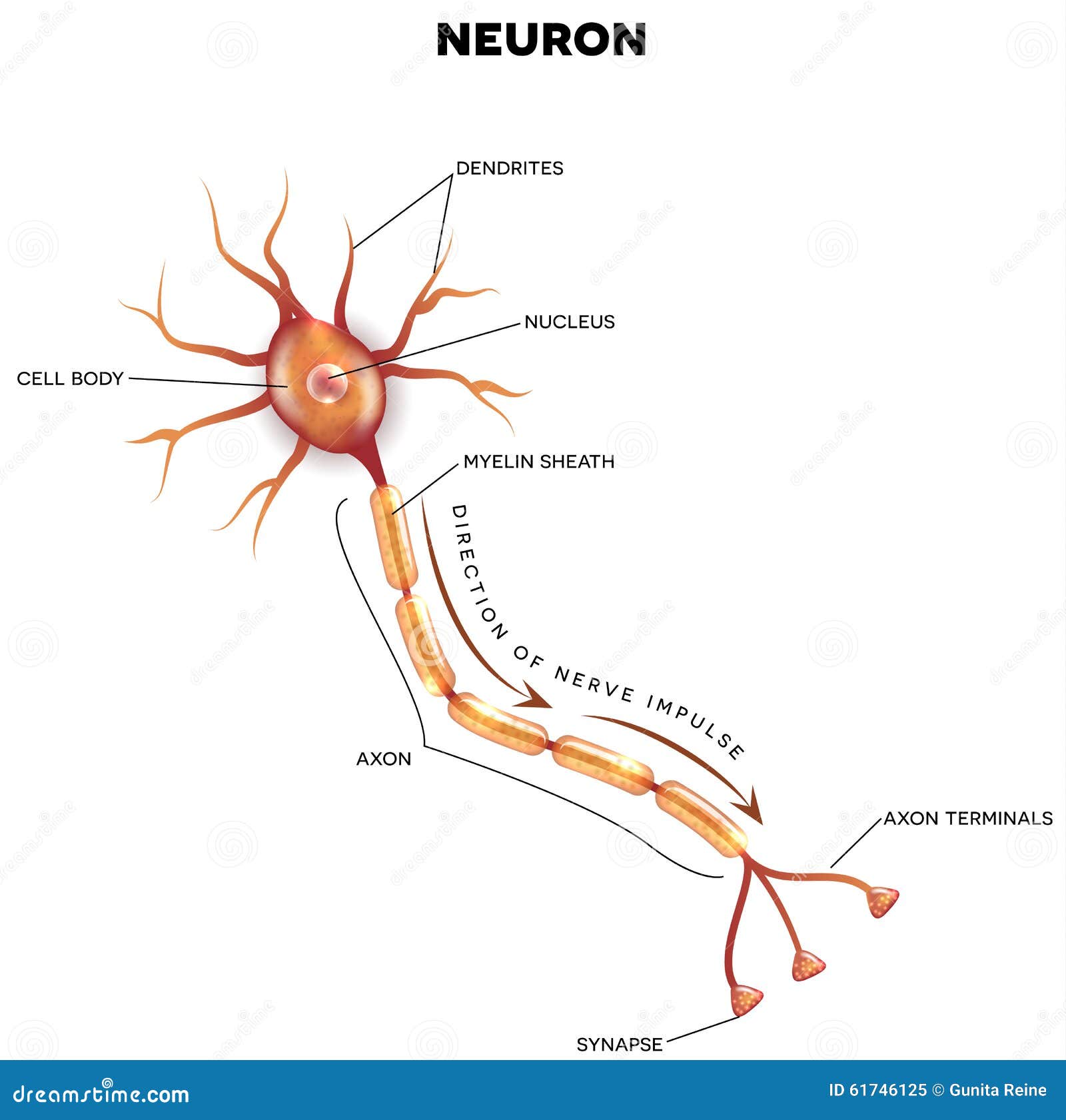
Most cells are 20 micrometers in diameter, which is just a fraction of the width of a hair. Neuron Anatomy Nerve Cell: Dendrites receive messages from other neurons. The message then moves through the axon to the other end of the neuron, then to the tips of the axon and then into the space between neurons.
Labeled Diagram of the Neuron Stock Vector Illustration of anatomical
The nervous system is a network of neurons whose main feature is to generate, modulate and transmit information between all the different parts of the human body. This property enables many important functions of the nervous system, such as regulation of vital body functions ( heartbeat, breathing, digestion), sensation and body movements.
